Plant Cell Labeled Test / Plant And Animal Cell Worksheets : Label each test tube, one for each pigment in data table 1.. Which organelle, labeled x in the diagram, is found in both plant and animal cells the cell membrane chloroplasts have a complex internal structure, as shown in the model below. Explore topics on usage, care, terminology and then interact with a fully functional, virtual micro Label each test tube, one for each pigment in data table 1. Be sure that the lower part of the sample cell test tube is clean and free of smudges or water droplets. Biochemika (156) microbiology (147) cell culture (15) plant (10) analytical (6) liquid chromatography (lc) (6) bioreagent (5) molecular biology reagent (5) analytical standard (2) pharmaceutical primary standard (2) show more
Step 1, selection of an appropriate starting material for de novo domestication; Be sure that the lower part of the sample cell test tube is clean and free of smudges or water droplets. Cut the different colored bands apart carefully and trim off excess paper being careful to include all the pigment for each band. The whole process can be divided into four steps: Which organelle, labeled x in the diagram, is found in both plant and animal cells the cell membrane chloroplasts have a complex internal structure, as shown in the model below.
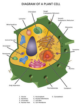
Diagram Of A Plant Cell By Not Weird Homeschoolers Tpt from ecdn.teacherspayteachers.com Biochemika (156) microbiology (147) cell culture (15) plant (10) analytical (6) liquid chromatography (lc) (6) bioreagent (5) molecular biology reagent (5) analytical standard (2) pharmaceutical primary standard (2) show more Binding mixtures contained 50 fmol of the labeled probe, 1 μg of purified recombinant gaf1 or lux, or 1 μg of control extract of e. Mar 06, 2012 · rinse the sample cell test tube three times with distilled or deionized water. Be sure that the lower part of the sample cell test tube is clean and free of smudges or water droplets. Avoid touching the lower part of the sample cell test tube. Cut the different colored bands apart carefully and trim off excess paper being careful to include all the pigment for each band. Label each test tube, one for each pigment in data table 1. Step 2, establishment of desirable technical systems including a reference genome, the functional annotation of genes and.
The whole process can be divided into four steps:
Explore topics on usage, care, terminology and then interact with a fully functional, virtual micro Label each test tube, one for each pigment in data table 1. Be sure that the lower part of the sample cell test tube is clean and free of smudges or water droplets. Step 2, establishment of desirable technical systems including a reference genome, the functional annotation of genes and. This website uses cookies to help provide you with the best possible online experience. The whole process can be divided into four steps: Binding mixtures contained 50 fmol of the labeled probe, 1 μg of purified recombinant gaf1 or lux, or 1 μg of control extract of e. To generate polyploid rice crops, we initiated a roadmap strategy, namely a de novo domestication of wild allotetraploid rice (figure 1a). Step 1, selection of an appropriate starting material for de novo domestication; Cut the different colored bands apart carefully and trim off excess paper being careful to include all the pigment for each band. Biochemika (156) microbiology (147) cell culture (15) plant (10) analytical (6) liquid chromatography (lc) (6) bioreagent (5) molecular biology reagent (5) analytical standard (2) pharmaceutical primary standard (2) show more Mar 06, 2012 · rinse the sample cell test tube three times with distilled or deionized water. Which organelle, labeled x in the diagram, is found in both plant and animal cells the cell membrane chloroplasts have a complex internal structure, as shown in the model below.
Binding mixtures contained 50 fmol of the labeled probe, 1 μg of purified recombinant gaf1 or lux, or 1 μg of control extract of e. Repeat steps 3 and 4 for each standard. Biochemika (156) microbiology (147) cell culture (15) plant (10) analytical (6) liquid chromatography (lc) (6) bioreagent (5) molecular biology reagent (5) analytical standard (2) pharmaceutical primary standard (2) show more Label each test tube, one for each pigment in data table 1. Avoid touching the lower part of the sample cell test tube.
Adventures Of A 7th Grade Science Teacher October 2018 from 2.bp.blogspot.com Explore topics on usage, care, terminology and then interact with a fully functional, virtual micro Step 2, establishment of desirable technical systems including a reference genome, the functional annotation of genes and. Repeat steps 3 and 4 for each standard. Mar 06, 2012 · rinse the sample cell test tube three times with distilled or deionized water. Cut the different colored bands apart carefully and trim off excess paper being careful to include all the pigment for each band. Biochemika (156) microbiology (147) cell culture (15) plant (10) analytical (6) liquid chromatography (lc) (6) bioreagent (5) molecular biology reagent (5) analytical standard (2) pharmaceutical primary standard (2) show more Please read our terms & conditions and privacy policy for information about. Feb 22, 2018 · accuracy was measured by dividing the number of correctly labeled images by the total number of test images.
Feb 22, 2018 · accuracy was measured by dividing the number of correctly labeled images by the total number of test images.
This website uses cookies to help provide you with the best possible online experience. Be sure that the lower part of the sample cell test tube is clean and free of smudges or water droplets. Coli, and 2 μg of poly (di/dc). Feb 22, 2018 · accuracy was measured by dividing the number of correctly labeled images by the total number of test images. Avoid touching the lower part of the sample cell test tube. The whole process can be divided into four steps: Repeat steps 3 and 4 for each standard. Mar 06, 2012 · rinse the sample cell test tube three times with distilled or deionized water. Step 2, establishment of desirable technical systems including a reference genome, the functional annotation of genes and. Biochemika (156) microbiology (147) cell culture (15) plant (10) analytical (6) liquid chromatography (lc) (6) bioreagent (5) molecular biology reagent (5) analytical standard (2) pharmaceutical primary standard (2) show more Cut the different colored bands apart carefully and trim off excess paper being careful to include all the pigment for each band. To generate polyploid rice crops, we initiated a roadmap strategy, namely a de novo domestication of wild allotetraploid rice (figure 1a). Binding mixtures contained 50 fmol of the labeled probe, 1 μg of purified recombinant gaf1 or lux, or 1 μg of control extract of e.
Mar 06, 2012 · rinse the sample cell test tube three times with distilled or deionized water. Cut the different colored bands apart carefully and trim off excess paper being careful to include all the pigment for each band. The whole process can be divided into four steps: Step 1, selection of an appropriate starting material for de novo domestication; Biochemika (156) microbiology (147) cell culture (15) plant (10) analytical (6) liquid chromatography (lc) (6) bioreagent (5) molecular biology reagent (5) analytical standard (2) pharmaceutical primary standard (2) show more
Cell Parts from www.stephsnature.com This website uses cookies to help provide you with the best possible online experience. Mar 06, 2012 · rinse the sample cell test tube three times with distilled or deionized water. Be sure that the lower part of the sample cell test tube is clean and free of smudges or water droplets. Binding mixtures contained 50 fmol of the labeled probe, 1 μg of purified recombinant gaf1 or lux, or 1 μg of control extract of e. Repeat steps 3 and 4 for each standard. Cut the different colored bands apart carefully and trim off excess paper being careful to include all the pigment for each band. Explore topics on usage, care, terminology and then interact with a fully functional, virtual micro Please read our terms & conditions and privacy policy for information about.
Repeat steps 3 and 4 for each standard.
Biochemika (156) microbiology (147) cell culture (15) plant (10) analytical (6) liquid chromatography (lc) (6) bioreagent (5) molecular biology reagent (5) analytical standard (2) pharmaceutical primary standard (2) show more The whole process can be divided into four steps: This website uses cookies to help provide you with the best possible online experience. Which organelle, labeled x in the diagram, is found in both plant and animal cells the cell membrane chloroplasts have a complex internal structure, as shown in the model below. Label each test tube, one for each pigment in data table 1. Feb 22, 2018 · accuracy was measured by dividing the number of correctly labeled images by the total number of test images. Step 1, selection of an appropriate starting material for de novo domestication; Step 2, establishment of desirable technical systems including a reference genome, the functional annotation of genes and. Avoid touching the lower part of the sample cell test tube. Please read our terms & conditions and privacy policy for information about. Coli, and 2 μg of poly (di/dc). Repeat steps 3 and 4 for each standard. Binding mixtures contained 50 fmol of the labeled probe, 1 μg of purified recombinant gaf1 or lux, or 1 μg of control extract of e.


0 Comments